
Advanced minimally invasive procedures for structural heart conditions
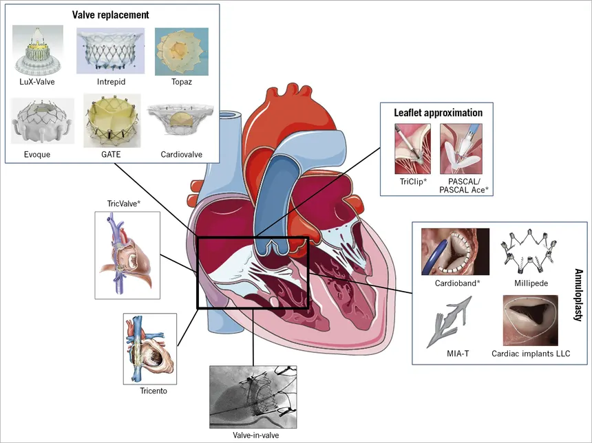
Tricuspid valve interventions are specialized treatments used to manage significant disease affecting the tricuspid valve, which controls blood flow between the right atrium and right ventricle of the heart. When the valve does not close properly, blood can flow backward into the right atrium, a condition known as tricuspid regurgitation. In other cases, the valve may become narrowed, restricting normal blood flow. Significant tricuspid valve disease can gradually affect the right side of the heart and may lead to symptoms such as breathlessness, fatigue and swelling of the legs or abdomen.
Modern advances in structural heart treatment have introduced minimally invasive tricuspid valve interventions for selected patients who may not be suitable for conventional open-heart surgery. Depending on the patient’s condition and valve anatomy, catheter-based procedures can be used to reduce valve leakage or, in appropriate cases, replace the diseased valve. These procedures aim to improve blood flow, reduce symptoms and support better heart function while avoiding the larger incisions associated with traditional surgery.
Tricuspid valve disease can develop because of several underlying conditions. Tricuspid regurgitation is frequently associated with changes in the right side of the heart caused by conditions such as pulmonary hypertension, left-sided heart disease, heart failure or enlargement of the right ventricle. In these situations, the valve may become stretched or displaced, preventing its leaflets from closing effectively.
Other causes include infection of the heart valve, congenital abnormalities, rheumatic heart disease, previous cardiac procedures and direct damage to the valve. Tricuspid valve disease can also occur as part of a broader pattern of heart valve disease. Identifying the underlying cause is important because treatment needs to address both the valve problem and any associated cardiac condition.
Tricuspid valve disease may remain unnoticed for a considerable period, particularly when the condition is mild. As valve dysfunction becomes more significant, pressure can build up within the veins and right side of the heart. Patients may experience increasing fatigue, reduced exercise tolerance, breathlessness and a feeling of weakness during everyday activities.
Fluid retention is another important feature of advanced tricuspid valve disease. Swelling of the feet, ankles or legs, abdominal swelling, discomfort caused by fluid accumulation and unexplained weight gain may occur. Some patients may also experience a feeling of fullness or reduced appetite. Because these symptoms can develop gradually, they may sometimes be mistaken for normal ageing or other medical problems.
Accurate diagnosis begins with a detailed cardiovascular assessment, including medical history, physical examination and evaluation of symptoms. Echocardiography is the primary investigation used to assess the tricuspid valve. It can provide important information about the degree of valve leakage or narrowing, the size and function of the right-sided heart chambers and the pressure within the heart and lungs.
Additional imaging and cardiac investigations may be recommended when an intervention is being considered. Transesophageal echocardiography can provide more detailed images of the valve, while cardiac CT or other investigations may help assess the anatomy and determine whether a minimally invasive procedure is appropriate. A comprehensive evaluation by a multidisciplinary heart valve team helps identify the severity of disease and select the safest and most effective treatment strategy.
Treatment depends on the severity of tricuspid valve disease, the patient’s symptoms, heart function, underlying cause and overall surgical risk. In patients with mild or stable disease, regular monitoring and appropriate medical treatment may be sufficient. Medications, particularly diuretics, may be used to control fluid retention and symptoms associated with heart failure, although medicines do not directly repair a structurally damaged tricuspid valve.
When tricuspid valve disease becomes severe and causes significant symptoms or affects heart function, an intervention may be considered. Traditional surgical repair or replacement remains an important treatment for selected patients. However, for carefully selected individuals who are at increased risk from conventional surgery, transcatheter tricuspid valve interventions offer a less invasive approach. Catheter-based edge-to-edge repair can bring the valve leaflets closer together to reduce regurgitation, while transcatheter valve replacement may be considered in selected patients with appropriate anatomy. The most suitable approach is determined after detailed imaging and individualized assessment.
Following a tricuspid valve intervention, patients are closely monitored to evaluate recovery, heart function and the performance of the treated valve. Medications may be prescribed or adjusted according to the patient’s individual cardiovascular condition. Managing associated conditions such as heart failure, high blood pressure, pulmonary hypertension or abnormal heart rhythms is also an important part of long-term care.
Regular follow-up is essential after treatment. Echocardiographic examinations may be performed periodically to assess the tricuspid valve, right-sided heart function and any remaining or recurrent valve leakage. Patients should follow their cardiologist’s recommendations regarding medications, physical activity, diet and other lifestyle measures. Maintaining consistent follow-up can help identify changes early and support long-term cardiovascular health.
Although modern transcatheter tricuspid valve procedures are designed to be less invasive than open-heart surgery, they still carry potential risks. Depending on the specific procedure and the patient’s health, complications may include bleeding or vascular problems at the catheter access site, infection, abnormal heart rhythms, blood clots, injury to cardiac structures or complications related to anesthesia or sedation. In some cases, the procedure may not sufficiently reduce valve leakage, or further treatment may be required.
The individual risk varies according to factors such as age, heart function, kidney function, pulmonary pressures, severity of valve disease and other medical conditions. Careful patient selection, advanced cardiac imaging and management by an experienced structural heart team are important in minimizing risks and achieving the best possible outcome.
Persistent breathlessness, unexplained fatigue, reduced exercise capacity, swelling of the legs or abdomen, rapid weight gain or a persistent feeling of abdominal fullness should be evaluated by a qualified cardiologist, particularly in patients with known heart or valve disease. Early diagnosis can help prevent progressive damage to the right side of the heart and allow appropriate treatment to be considered at the right time.
Patients who have already been diagnosed with tricuspid valve disease should maintain regular cardiovascular follow-up even if their symptoms are mild. Sudden or rapidly worsening breathlessness, severe swelling, chest discomfort, fainting or significant deterioration in exercise tolerance requires prompt medical assessment. With appropriate evaluation and advances in minimally invasive structural heart procedures, many patients can now be considered for individualized treatment options that were previously unavailable.
Explore More: Atrial Septal Defect (ASD) Closure

Providing advanced, compassionate and evidence-based cardiac care with a focus on interventional cardiology, structural heart procedures and comprehensive heart health.
9:00 AM – 6:00 PM (By Appointment Only)
No WhatsApp Number Found!
WhatsApp us