
Advanced minimally invasive procedures for structural heart conditions
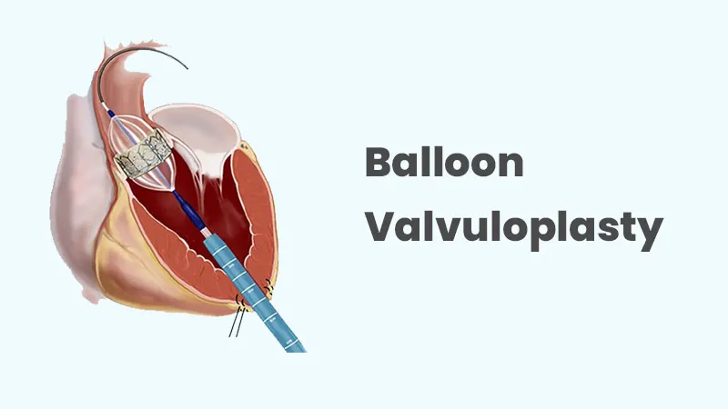
Balloon valvuloplasty, also known as balloon valvotomy, is a minimally invasive cardiac procedure used to improve blood flow through a narrowed heart valve. It is most commonly performed for certain patients with significant narrowing of the mitral valve or pulmonary valve and, in selected circumstances, other heart valves. Valve narrowing, known as stenosis, can make it difficult for blood to move efficiently through the heart and may gradually increase the workload on the heart muscle.
During balloon valvuloplasty, a thin catheter with a specially designed balloon at its tip is guided through a blood vessel to the affected heart valve. Once correctly positioned, the balloon is briefly inflated to separate or stretch the narrowed valve opening. The balloon is then deflated and removed, allowing blood to flow more freely through the valve. As a catheter-based treatment, balloon valvuloplasty generally avoids the need for open-heart surgery and may provide symptom relief and improved circulation in appropriately selected patients.
Heart valve narrowing can develop for different reasons depending on the valve involved. Rheumatic heart disease is an important cause of mitral stenosis, particularly in regions where rheumatic fever remains prevalent. The valve may become thickened, stiff or fused over time, gradually restricting the opening through which blood passes.
Pulmonary valve stenosis is often congenital, meaning the valve develops abnormally before birth. In other patients, valve narrowing may develop because of age-related changes, calcium deposits or other structural abnormalities. Identifying the underlying cause and understanding the anatomy of the affected valve are essential when determining whether balloon valvuloplasty is an appropriate treatment.
Mild valve stenosis may cause few or no symptoms for many years. As the narrowing becomes more significant, the heart has to work harder to push blood through the restricted valve. Patients may gradually develop shortness of breath, particularly during physical activity, along with fatigue and reduced exercise tolerance.
Depending on the valve affected and the severity of the narrowing, symptoms may also include chest discomfort, dizziness, fainting, palpitations or swelling of the legs and feet. Some patients may experience symptoms only during exertion, while more advanced valve disease can cause breathlessness during routine activities or even at rest. Persistent or worsening symptoms should be evaluated by a cardiologist.
The evaluation of valve stenosis generally begins with a detailed medical history, physical examination and assessment of cardiovascular symptoms. A heart murmur detected during examination may provide an important clue that further investigation is required. Echocardiography is the primary imaging test used to evaluate heart valve disease because it can assess the structure and movement of the valve, the severity of narrowing and the effect of the condition on the heart chambers.
Additional investigations may be recommended depending on the patient’s condition and the valve being considered for treatment. An electrocardiogram can evaluate heart rhythm and electrical activity, while chest imaging and other cardiac tests may provide additional information. Transesophageal echocardiography, cardiac catheterization or other advanced imaging may be used when detailed anatomical information is needed before balloon valvuloplasty.
Treatment depends on the severity of valve stenosis, the patient’s symptoms, heart function, valve anatomy and overall health. Patients with mild or stable disease may be monitored regularly, while medications may be prescribed to manage associated symptoms or conditions. Medications, however, generally cannot remove the physical narrowing of a severely stenotic valve.
For carefully selected patients, balloon valvuloplasty can provide a minimally invasive way to enlarge the narrowed valve opening. The procedure involves advancing a catheter, usually through a blood vessel in the groin, toward the affected valve. Once positioned, the balloon is inflated for a short period to separate fused valve tissue or stretch the narrowed opening. In patients who are not suitable candidates for balloon treatment, surgical valve repair or replacement, or other transcatheter valve procedures, may be considered depending on the individual valve condition. Treatment decisions should be based on detailed cardiac imaging and assessment by an experienced heart team.
After balloon valvuloplasty, patients are monitored to assess heart rhythm, blood pressure, valve function and the catheter access site. The length of hospital observation depends on the valve treated, the patient’s overall health and the complexity of the procedure. Some patients can return to normal activities relatively quickly, although strenuous physical activity may need to be limited temporarily according to medical advice.
Follow-up appointments are important to assess the long-term effectiveness of the procedure. Echocardiography may be performed to evaluate the treated valve and monitor for any remaining or recurrent narrowing. Patients should continue prescribed medications and maintain appropriate control of blood pressure, diabetes and other cardiovascular risk factors. Regular cardiac follow-up is particularly important because valve narrowing can sometimes recur or progress over time.
Balloon valvuloplasty is generally considered a minimally invasive procedure, but it carries potential risks. These may include bleeding or bruising at the catheter insertion site, blood-vessel injury, abnormal heart rhythms, infection or reactions related to medications or contrast material when used. The risk profile varies depending on the valve being treated and the patient’s individual health.
In some cases, the procedure may not provide sufficient enlargement of the valve opening, or the valve may gradually become narrowed again. Balloon treatment can also occasionally cause or worsen valve leakage, known as regurgitation. Rare but serious complications can include blood clots, significant bleeding, damage to cardiac structures, stroke or other cardiovascular events. Careful patient selection and detailed pre-procedure assessment help minimize these risks.
Patients experiencing unexplained breathlessness, persistent fatigue, reduced exercise capacity, chest discomfort, dizziness, fainting or recurrent palpitations should consider a cardiovascular evaluation, particularly if they have previously been diagnosed with heart valve disease. Early diagnosis can help determine the severity of valve narrowing and whether regular monitoring or an intervention may be appropriate.
Patients who have undergone balloon valvuloplasty should attend scheduled follow-up appointments and report any new or worsening symptoms to their cardiologist. Sudden severe breathlessness, chest pain, fainting, significant bleeding or symptoms suggestive of a stroke require immediate medical attention. Timely evaluation allows appropriate treatment to be provided before valve disease leads to more significant strain on the heart.
Explore More: Mitral Valvuloplasty

Providing advanced, compassionate and evidence-based cardiac care with a focus on interventional cardiology, structural heart procedures and comprehensive heart health.
9:00 AM – 6:00 PM (By Appointment Only)
No WhatsApp Number Found!
WhatsApp us