
Advanced minimally invasive procedures for structural heart conditions
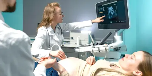
Advanced Echocardiography is a specialized form of cardiac ultrasound that provides detailed information about the structure and function of the heart. Using sophisticated imaging techniques, it allows a cardiologist to assess the heart chambers, valves, muscle, blood flow and overall pumping function with greater precision than a routine echocardiogram. Depending on the clinical requirement, advanced echocardiographic assessment may include three-dimensional echocardiography, Doppler imaging, strain imaging, contrast echocardiography or transesophageal echocardiography.
This advanced cardiac imaging plays an important role in diagnosing and monitoring a wide range of cardiovascular conditions. It can help identify abnormalities that may not be fully characterized by a standard echocardiogram and can provide valuable information when planning procedures such as valve repair, valve replacement or structural heart interventions. Advanced Echocardiography is a non-invasive or minimally invasive diagnostic approach, depending on the technique used, and enables personalized cardiac evaluation based on the patient’s symptoms and medical history.
Advanced Echocardiography is not performed because of a single disease or cause. Instead, it is recommended when a patient has symptoms, clinical findings or a known heart condition that requires more detailed assessment. Breathlessness, chest discomfort, unexplained fatigue, palpitations, swelling of the legs or reduced exercise tolerance may lead a cardiologist to recommend detailed cardiac imaging.
It may also be used when a routine echocardiogram identifies an abnormality that requires further evaluation. Patients with suspected or known heart valve disease, cardiomyopathy, heart failure, congenital heart defects, pulmonary hypertension or abnormalities of the heart chambers may benefit from advanced echocardiographic assessment. The investigation can also be particularly useful before and after structural heart procedures to evaluate treatment results and monitor cardiac function.
Advanced Echocardiography itself does not have symptoms because it is a diagnostic investigation. The symptoms that lead to an advanced echocardiographic evaluation usually arise from an underlying cardiovascular condition. Patients may experience shortness of breath, persistent tiredness, dizziness, fainting, palpitations, chest discomfort or difficulty performing routine physical activities.
Some heart conditions may cause few or no noticeable symptoms in their early stages. In these situations, abnormal findings during a routine health examination, an unusual heart sound or changes seen on an electrocardiogram may prompt further cardiac imaging. Advanced Echocardiography can help investigate these findings and determine whether there is an underlying structural or functional abnormality.
Advanced Echocardiography uses high-frequency sound waves to create detailed moving images of the heart. Doppler techniques can evaluate the direction and speed of blood flow through the heart and across the valves, while advanced measurements can provide information about the movement and performance of the heart muscle. Three-dimensional imaging may provide a more detailed view of cardiac structures and can be especially useful when assessing complex valve or structural abnormalities.
Depending on the clinical situation, a transthoracic echocardiogram may be sufficient, while some patients may require a transesophageal echocardiogram. In transesophageal echocardiography, a specialized ultrasound probe is gently positioned in the oesophagus to obtain highly detailed images of structures that may be difficult to visualize from outside the chest. Advanced techniques such as myocardial strain imaging can also help detect subtle changes in heart muscle function that may not be apparent from conventional measurements.
Advanced Echocardiography is primarily a diagnostic and monitoring procedure rather than a treatment itself. The findings help the cardiologist determine the underlying cardiac condition and select the most appropriate treatment strategy. Depending on the results, management may include medication, lifestyle modification, regular monitoring or referral for an appropriate cardiac intervention.
In patients with significant heart valve disease or structural heart conditions, detailed echocardiographic imaging can assist in determining whether procedures such as valve repair, valve replacement or catheter-based interventions may be appropriate. Advanced imaging can also be used during the planning and follow-up of certain cardiac procedures, helping the medical team assess anatomy, treatment response and changes in heart function.
Because Advanced Echocardiography is primarily an imaging investigation, most patients can return to their usual activities immediately after a standard transthoracic examination. If a transesophageal echocardiogram or another specialized technique involving sedation has been performed, additional observation and temporary restrictions may be required according to the medical team’s instructions.
The most important aspect of follow-up is understanding and acting on the results. Depending on the findings, the cardiologist may recommend medication, additional investigations, periodic echocardiographic monitoring or further treatment. Patients with diagnosed heart conditions should attend scheduled follow-up appointments so that changes in heart function or disease progression can be identified at an appropriate stage.
Standard transthoracic echocardiography is considered a safe diagnostic test and does not involve ionizing radiation. It is generally well tolerated and has very few risks. Advanced imaging techniques may have additional considerations depending on how the examination is performed.
Transesophageal echocardiography is generally safe when performed by an experienced medical team, but it can occasionally cause temporary throat discomfort or irritation. Rare complications can include injury to the oesophagus, bleeding, breathing difficulties or reactions related to sedation. Contrast echocardiography, when required, may also carry a small risk of an allergic or other adverse reaction. The medical team will assess the patient’s health and explain any relevant precautions before a specialized examination.
Patients should consider a cardiac evaluation if they develop persistent or unexplained breathlessness, chest discomfort, palpitations, dizziness, fainting, unusual fatigue or a noticeable decline in exercise capacity. A cardiologist may recommend an echocardiogram when these symptoms suggest a possible structural or functional heart problem.
Advanced Echocardiography may be particularly valuable when a previous cardiac test has shown an abnormality or when a known heart condition requires detailed assessment. Patients already being treated for heart valve disease, heart failure, cardiomyopathy or other structural heart conditions should follow their recommended monitoring schedule. Sudden severe chest pain, severe difficulty breathing, fainting or symptoms suggestive of a stroke require immediate emergency medical attention rather than waiting for a routine cardiac appointment.
Explore More: Transesophageal Echocardiography (TEE)

Providing advanced, compassionate and evidence-based cardiac care with a focus on interventional cardiology, structural heart procedures and comprehensive heart health.
9:00 AM – 6:00 PM (By Appointment Only)
No WhatsApp Number Found!
WhatsApp us