
Advanced minimally invasive procedures for structural heart conditions
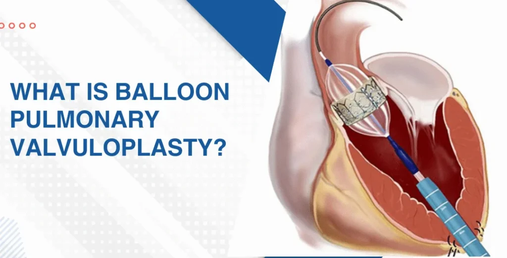
Pulmonary valvuloplasty, also known as balloon pulmonary valvuloplasty, is a minimally invasive catheter-based procedure used to treat narrowing of the pulmonary valve, a condition known as pulmonary valve stenosis. The pulmonary valve controls blood flow from the right ventricle of the heart into the pulmonary artery, which carries blood toward the lungs. When this valve becomes narrowed, the right side of the heart has to work harder to pump blood through the narrowed opening. Pulmonary valvuloplasty helps widen the valve opening and improve blood flow from the heart to the lungs.
During the procedure, a thin catheter with a small balloon at its tip is guided through a blood vessel, usually from the groin, toward the heart. Once the balloon is positioned across the narrowed pulmonary valve, it is briefly inflated to separate the valve leaflets and enlarge the opening. The balloon is then deflated and removed. Because the procedure does not require open-heart surgery, recovery is generally quicker, making it an important treatment option for appropriately selected patients, including children and adults with significant pulmonary valve stenosis.
Pulmonary valve stenosis is most commonly a congenital heart condition, meaning the pulmonary valve develops abnormally before birth. The valve may have thickened or fused leaflets that do not open normally, restricting blood flow from the right ventricle to the lungs. In many patients, the exact reason for the abnormal development is not known.
Less commonly, pulmonary valve narrowing can develop later in life due to conditions that affect the heart valves. Certain congenital heart diseases may also occur alongside pulmonary stenosis. The severity of narrowing can vary considerably, and some patients may have mild disease that only requires monitoring, while significant stenosis may eventually require an intervention such as pulmonary valvuloplasty.
Mild pulmonary valve stenosis may not cause noticeable symptoms and can sometimes remain undiagnosed for years. As the narrowing becomes more severe, the right side of the heart must generate greater pressure to push blood through the valve. Patients may experience shortness of breath, especially during physical activity, fatigue, reduced exercise tolerance, dizziness or fainting.
Some individuals may experience chest discomfort or a sensation of rapid or irregular heartbeat. In children, significant pulmonary stenosis may sometimes be associated with poor exercise tolerance, difficulty feeding, inadequate weight gain or other signs of reduced cardiovascular function. The severity and combination of symptoms depend on the degree of valve narrowing and the overall condition of the heart.
Diagnosis of pulmonary valve stenosis usually begins with a medical history, physical examination and cardiac evaluation. A heart murmur may be detected during a routine examination and can lead to further testing. Echocardiography is the primary investigation used to assess the pulmonary valve, measure the degree of narrowing and evaluate blood flow through the valve. It can also provide information about the size and function of the right ventricle.
An electrocardiogram may be performed to assess the heart’s electrical activity, while additional imaging or cardiac catheterization may be recommended in selected cases. Cardiac catheterization can provide direct measurements of pressure within the heart and pulmonary circulation and may be performed as part of the evaluation or during the valvuloplasty procedure itself. A detailed assessment helps determine the severity of pulmonary stenosis and whether intervention is appropriate.
Treatment depends on the severity of pulmonary valve stenosis, the patient’s symptoms, the pressure within the right side of the heart and the overall condition of the pulmonary valve. Patients with mild narrowing and no significant symptoms may only require regular cardiac examinations and echocardiographic monitoring. Medications generally do not correct the physical narrowing of the valve but may be used to manage associated symptoms or other cardiac conditions when necessary.
For significant valvar pulmonary stenosis, balloon pulmonary valvuloplasty is often an effective minimally invasive treatment. A catheter-mounted balloon is positioned across the narrowed valve and inflated to widen the valve opening, reducing the obstruction and allowing blood to flow more freely toward the lungs. In patients whose valve anatomy is unsuitable for balloon treatment, or when significant valve dysfunction is present, surgical pulmonary valve repair or replacement may be considered. The most suitable approach is determined after careful assessment of the valve anatomy and the patient’s individual needs.
After pulmonary valvuloplasty, patients are monitored for changes in heart rhythm, blood pressure, valve function and the catheter access site. The duration of hospital observation varies depending on the patient’s age, overall health and response to the procedure. Most appropriately selected patients can return to normal activities gradually, according to their cardiologist’s recommendations.
Regular follow-up is important to monitor the treated pulmonary valve and the function of the right side of the heart. Echocardiography may be performed periodically to assess blood flow across the valve and identify any residual or recurrent narrowing or pulmonary valve leakage. Maintaining scheduled appointments and following medication and activity recommendations can support a safe recovery and long-term heart health.
Pulmonary valvuloplasty is generally considered a safe and effective procedure, but as with any cardiac intervention, it carries potential risks. These may include bleeding or bruising at the catheter insertion site, blood-vessel complications, infection, abnormal heart rhythms or temporary changes in blood pressure. Some patients may develop pulmonary valve regurgitation, in which the valve allows some blood to flow backward after the procedure.
In some cases, the valve may remain partially narrowed or may become narrowed again over time, requiring continued monitoring or further treatment. Rarely, more serious complications involving the heart or blood vessels can occur. The individual risk depends on factors such as the severity of pulmonary stenosis, valve anatomy, age and overall health. Careful evaluation and experienced procedural care help minimize these risks.
Medical evaluation is recommended if you or your child develops unexplained shortness of breath, excessive fatigue, reduced exercise tolerance, dizziness, fainting, chest discomfort or persistent palpitations. A heart murmur identified during a routine examination should also be evaluated, particularly when there are other signs or symptoms of possible heart disease.
Patients with known pulmonary valve stenosis should maintain regular follow-up with a cardiologist to monitor the severity of the condition and the function of the right side of the heart. If symptoms become progressively worse or significantly limit daily activities, further evaluation may be necessary to determine whether pulmonary valvuloplasty or another form of treatment is appropriate. Sudden severe breathlessness, fainting, significant chest pain or other acute symptoms require prompt medical attention.
Explore More: Transcatheter Valve-in-Valve Procedures

Providing advanced, compassionate and evidence-based cardiac care with a focus on interventional cardiology, structural heart procedures and comprehensive heart health.
9:00 AM – 6:00 PM (By Appointment Only)
No WhatsApp Number Found!
WhatsApp us