
Advanced minimally invasive procedures for structural heart conditions
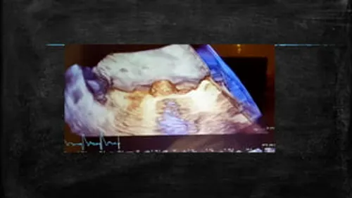
3D Echocardiography is an advanced cardiac imaging technique that creates detailed three-dimensional images of the heart in real time. Unlike conventional two-dimensional echocardiography, which provides flat images from selected views, 3D echocardiography allows the heart’s structures to be visualized from multiple angles and reconstructed as a three-dimensional image. This can provide valuable information about the size, shape and movement of the heart chambers, valves and other important cardiac structures.
3D echocardiography is particularly useful when a more detailed understanding of heart anatomy is required. It can assist cardiologists in evaluating complex valve disorders, congenital heart abnormalities, heart chamber function and other structural heart conditions. The technique may also play an important role in planning and guiding certain cardiac procedures, helping the medical team make more precise treatment decisions based on detailed anatomical information.
3D echocardiography itself does not treat or cause a disease; rather, it is used to investigate and evaluate various cardiac conditions. A doctor may recommend this advanced imaging technique when there is concern about abnormalities involving the heart valves, chambers, walls or surrounding structures. Conditions such as mitral valve disease, aortic valve disorders, congenital heart defects and certain forms of heart failure may require detailed three-dimensional assessment.
It may also be recommended when conventional echocardiography does not provide enough information to fully understand the heart’s anatomy. In patients being evaluated for procedures such as valve repair, valve replacement or structural heart interventions, 3D imaging can provide a more comprehensive view of the relevant cardiac structures and help support individualized treatment planning.
3D echocardiography does not produce symptoms because it is a diagnostic procedure. Instead, it may be recommended when a patient has symptoms that could indicate an underlying heart condition. These symptoms may include unexplained breathlessness, chest discomfort, fatigue, reduced exercise tolerance, dizziness, fainting or a sensation of rapid or irregular heartbeat.
Some structural heart conditions may cause few or no symptoms during their early stages and may be discovered during a routine examination or conventional echocardiogram. When further anatomical information is needed, 3D echocardiography can provide additional detail and help determine whether a significant cardiac abnormality is present.
3D echocardiography uses ultrasound waves to produce detailed images of the heart without exposing the patient to radiation. Depending on the clinical requirement, it can be performed using a transthoracic approach, in which the ultrasound probe is placed on the chest, or through transesophageal echocardiography, where a specialized probe is guided into the esophagus to obtain closer images of the heart.
The examination can provide detailed information about the structure and function of the heart, including valve anatomy, chamber dimensions, movement of the heart walls and blood flow-related abnormalities. Three-dimensional imaging can be particularly valuable for assessing the mitral valve and other complex structures because it allows the cardiologist to examine anatomy from different perspectives. The findings are interpreted alongside the patient’s symptoms, medical history and other cardiac investigations to establish an accurate diagnosis.
3D echocardiography is a diagnostic and imaging procedure rather than a treatment itself. Its primary role is to provide detailed information that helps the cardiologist determine the most appropriate management for the underlying heart condition. The treatment recommended after the examination depends entirely on what the imaging reveals and how significantly the condition is affecting the patient’s heart function.
Depending on the diagnosis, treatment may involve regular monitoring, medications, lifestyle and risk-factor management, minimally invasive cardiac procedures or conventional surgery. In patients with significant heart valve disease, 3D echocardiography can also assist in assessing whether a patient may be suitable for procedures such as valve repair or transcatheter interventions. Detailed imaging allows treatment decisions to be tailored more precisely to each patient’s cardiac anatomy and clinical needs.
There is generally no prolonged recovery period after a routine transthoracic 3D echocardiogram, and most patients can return to their normal activities immediately after the examination. If a transesophageal 3D echocardiogram is performed, the patient may require a short period of observation following the procedure, particularly because sedation or throat-numbing medication may be used. Specific instructions will be provided by the medical team.
When 3D echocardiography is performed as part of the evaluation or follow-up of a cardiac condition, ongoing care depends on the underlying diagnosis. Patients may require prescribed medications, periodic echocardiographic monitoring, lifestyle modifications or additional cardiac procedures. Attending scheduled follow-up appointments allows the cardiologist to monitor changes in heart function and adjust treatment when necessary.
Transthoracic 3D echocardiography is generally considered a safe and non-invasive examination and does not involve radiation. It typically has very few risks. Some patients may experience mild pressure from the ultrasound probe against the chest, but the examination is usually well tolerated.
Transesophageal 3D echocardiography is more invasive and carries additional considerations because the imaging probe is passed into the esophagus. Temporary throat discomfort, gagging or minor irritation may occur. Rarely, complications such as bleeding, injury to the esophagus, breathing difficulties or reactions to sedation can occur. The medical team carefully evaluates the patient’s health and medical history before recommending this type of examination.
You should consider a cardiac evaluation if you experience persistent or unexplained breathlessness, chest discomfort, unusual fatigue, dizziness, fainting, palpitations or a noticeable decline in your ability to perform normal physical activities. A doctor may recommend an echocardiogram when these symptoms suggest a possible structural or functional heart problem.
3D echocardiography may be particularly valuable when a more detailed assessment is needed after an initial cardiac examination or when planning treatment for a structural heart condition. Regular cardiac follow-up is also important for patients who have already been diagnosed with heart valve disease, congenital heart abnormalities, heart failure or other cardiovascular conditions. If symptoms such as severe chest pain, sudden breathlessness or loss of consciousness occur suddenly, immediate medical attention should be sought.
Explore More: Stress Echocardiography

Providing advanced, compassionate and evidence-based cardiac care with a focus on interventional cardiology, structural heart procedures and comprehensive heart health.
9:00 AM – 6:00 PM (By Appointment Only)
No WhatsApp Number Found!
WhatsApp us