
Advanced minimally invasive procedures for structural heart conditions
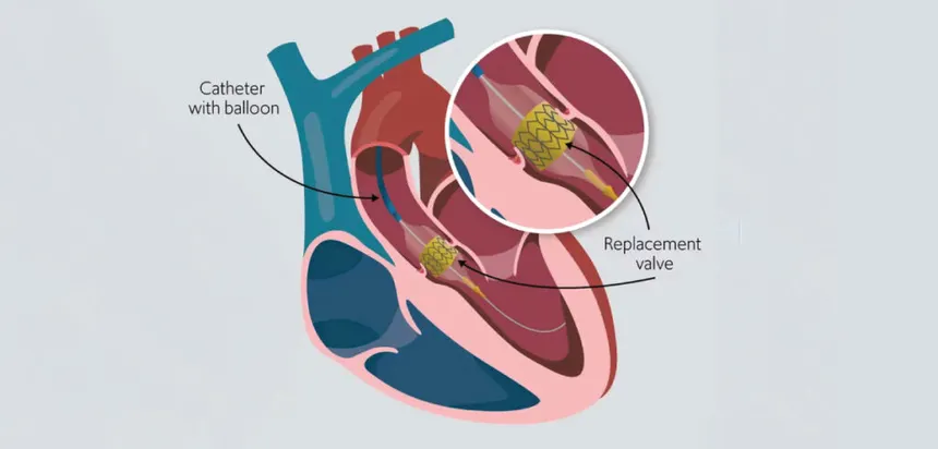
Transcatheter Valve-in-Valve (ViV) procedures are minimally invasive treatments used to manage a previously implanted surgical heart valve that has become narrowed, stiff or leaky over time. Biological or tissue-based replacement valves can gradually deteriorate, leading to a condition known as bioprosthetic valve dysfunction. Traditionally, treatment of a failing replacement valve may require another open-heart surgery. A transcatheter valve-in-valve procedure provides a less invasive alternative for carefully selected patients by placing a new transcatheter heart valve inside the existing surgical valve.
During the procedure, a specially designed replacement valve is delivered through a thin catheter, commonly through a blood vessel in the groin, and positioned within the old bioprosthetic valve. Once expanded, the new valve takes over the function of the failing valve and helps restore more effective blood flow. Depending on the location of the previous surgical valve, valve-in-valve treatment may be performed for selected aortic, mitral, pulmonary or tricuspid bioprosthetic valves. The appropriate approach depends on the patient’s valve anatomy, previous surgery, symptoms and overall health.
The need for a valve-in-valve procedure generally develops because a previously implanted biological heart valve has deteriorated over time. Tissue valves are designed to provide long-term valve function, but they can gradually develop structural changes, including calcium deposits, thickening, tearing or loss of normal flexibility. This deterioration can cause the valve to become narrowed, known as stenosis, or allow blood to leak backward, known as regurgitation.
The likelihood and timing of bioprosthetic valve deterioration vary between individuals and depend on factors such as the type and position of the original valve, age at implantation, underlying heart disease and other medical conditions. Regular follow-up after valve replacement is important because structural valve deterioration can sometimes progress before significant symptoms become apparent.
A failing biological heart valve can produce symptoms similar to those caused by other forms of valve disease. Patients may experience increasing breathlessness, tiredness, reduced ability to exercise, dizziness or swelling of the legs and ankles. Some people may also notice chest discomfort, palpitations or difficulty performing everyday activities that were previously comfortable.
Symptoms often develop gradually as the replacement valve becomes less effective. Persistent or worsening breathlessness, particularly during exertion or while lying down, may indicate that the heart is working harder to maintain adequate blood circulation. Early evaluation is important because timely treatment may prevent further deterioration of heart function.
Evaluation of a failing surgical valve begins with a detailed medical history, physical examination and cardiac assessment. Echocardiography is one of the most important investigations because it can assess the movement of the prosthetic valve, measure blood flow across it and identify narrowing or leakage. It can also provide information about the size and function of the heart chambers and the pressure within the heart and lungs.
Additional imaging may be required when a valve-in-valve procedure is being considered. Transesophageal echocardiography can provide detailed views of the valve and surrounding structures, while cardiac CT can help assess the size and anatomy of the existing prosthetic valve and determine the feasibility and safety of placing a new valve within it. Electrocardiography and other cardiac investigations may also be performed. A comprehensive assessment by a specialised heart valve team is essential for determining whether valve-in-valve treatment is appropriate.
Treatment depends on the severity of prosthetic valve dysfunction, the patient’s symptoms, heart function, age, overall health and the characteristics of the existing valve. Patients with mild valve deterioration and few or no symptoms may be monitored regularly with clinical examinations and echocardiography. Medications may be used to manage associated conditions and symptoms, although they do not correct a structurally failing prosthetic valve.
For selected patients with a deteriorated surgical bioprosthetic valve, a transcatheter valve-in-valve procedure can provide a minimally invasive treatment option. A replacement transcatheter valve is delivered through a catheter and positioned inside the existing surgical valve, where it expands and begins functioning as the new valve. This approach can avoid the need for repeat open-heart surgery in appropriately selected patients. However, valve-in-valve treatment is not suitable for every patient, and factors such as the size and type of the previous valve, its position, coronary artery anatomy and the risk of obstruction must be carefully assessed before treatment.
After a transcatheter valve-in-valve procedure, patients are closely monitored to assess the new valve’s function, heart rhythm and the catheter access site. Medication may be prescribed to reduce the risk of blood clots or manage other cardiovascular conditions, depending on the type of valve procedure and the patient’s individual medical history. Patients should take prescribed medications consistently and attend all recommended follow-up appointments.
Recovery is generally faster than after conventional repeat open-heart valve surgery, although the recovery period varies from person to person. Follow-up echocardiography is important to evaluate the performance of the new valve and monitor heart function over time. Patients should gradually return to physical activity according to their cardiologist’s recommendations and continue managing important cardiovascular risk factors such as high blood pressure, diabetes and high cholesterol.
Although transcatheter valve-in-valve procedures are minimally invasive, they are still significant cardiac interventions and can involve potential complications. These may include bleeding or damage to the blood vessel used for catheter access, infection, abnormal heart rhythms, blood clots, stroke or problems involving the newly implanted valve. Some patients may develop a residual pressure gradient across the new valve, particularly when a smaller surgical valve is being treated.
Depending on the location and anatomy of the existing valve, there may also be a risk of obstruction of nearby cardiac structures or coronary arteries. In some situations, a valve-in-valve procedure may not provide the desired result or may not be technically appropriate, making repeat surgery a more suitable option. Careful imaging, procedural planning and evaluation by an experienced multidisciplinary heart valve team are important for minimizing these risks.
Patients who have previously undergone surgical heart valve replacement should maintain regular cardiac follow-up even when they feel well. New or worsening breathlessness, fatigue, reduced exercise tolerance, dizziness, chest discomfort, palpitations or swelling of the legs should be evaluated, particularly in individuals with a known biological prosthetic valve.
If investigations show that a previously implanted valve is becoming significantly narrowed or leaky, a specialist assessment can help determine the most appropriate treatment. Depending on the patient’s condition and valve anatomy, continued monitoring, medical management, transcatheter valve-in-valve treatment or repeat surgical valve replacement may be considered. Sudden severe breathlessness, fainting, chest pain, significant weakness or other acute symptoms require immediate medical attention.
Explore More: Pulmonary Hypertension Evaluation

Providing advanced, compassionate and evidence-based cardiac care with a focus on interventional cardiology, structural heart procedures and comprehensive heart health.
9:00 AM – 6:00 PM (By Appointment Only)
No WhatsApp Number Found!
WhatsApp us