
Advanced cardiac procedures
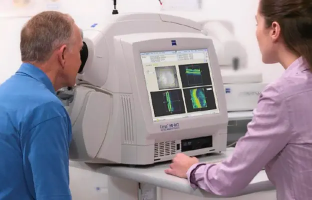
Optical Coherence Tomography (OCT) is a modern, non-invasive eye imaging technology that provides highly detailed, cross-sectional images of the retina, macula, optic nerve and other structures of the eye. It uses light waves to capture microscopic images of ocular tissues, allowing an eye specialist to identify subtle structural changes that may not be clearly visible during a routine eye examination. OCT is widely used for evaluating and monitoring conditions such as glaucoma, diabetic retinal disease, age-related macular degeneration, macular oedema, macular holes and other retinal disorders. Because the examination is quick and does not require direct contact with the eye, it is generally comfortable for patients.
OCT itself does not have a cause because it is a diagnostic imaging procedure rather than a disease. Instead, an ophthalmologist may recommend an OCT scan when there is a concern about changes affecting the retina, macula, optic nerve or other structures of the eye. Conditions associated with diabetes, glaucoma, age-related retinal changes, retinal swelling, macular disorders and certain optic nerve problems may require detailed OCT imaging. OCT may also be advised as part of routine monitoring when a patient has an existing eye condition or is undergoing treatment, helping the doctor compare changes over time.
The symptoms that may lead to an OCT examination depend on the underlying eye condition. Patients may experience blurred or distorted vision, difficulty reading, reduced central vision, blind spots, changes in colour perception or gradual loss of visual clarity. Some conditions, particularly glaucoma, may cause few or no noticeable symptoms in their early stages, which is why appropriate eye examinations and imaging can be important even when vision appears normal. An OCT scan can help detect structural changes that may occur before significant symptoms become apparent.
An OCT examination is performed using a specialised imaging device that scans the eye with light and produces detailed images of its internal structures. The patient is asked to look at a target while the machine captures the required images, and the process generally takes only a short time. Depending on the clinical concern, the doctor may examine retinal thickness, individual retinal layers, the macula, retinal nerve fibre layer or optic nerve. OCT findings are interpreted alongside the patient’s symptoms, medical history and other eye examinations because an OCT image alone may not always provide a complete diagnosis. In some cases, additional investigations may be recommended to understand the condition more accurately.
OCT is a diagnostic and monitoring tool, so it does not directly treat an eye condition. Instead, the results can help the ophthalmologist determine the most appropriate treatment according to the underlying diagnosis. Depending on the condition, management may involve prescription eye drops, injections, laser treatment, medication, surgery or regular observation. For example, OCT can be particularly valuable when monitoring retinal swelling or other retinal abnormalities and assessing how the eye responds to treatment over time. The recommended treatment will depend on the patient’s individual condition, severity and overall eye health.
Post-treatment care depends on the eye condition and the treatment performed rather than on the OCT scan itself. Patients should follow their ophthalmologist’s instructions regarding prescribed medicines, eye drops, follow-up appointments and activity restrictions. When OCT is being used for ongoing monitoring, attending scheduled scans can help the doctor compare retinal or optic nerve changes over time and assess whether the current treatment remains effective. Patients should also report any new or worsening changes in vision promptly rather than waiting for their next scheduled appointment.
OCT is generally considered a non-invasive and well-tolerated imaging examination, and it does not involve needles or direct contact with the eye. However, the quality of the scan can sometimes be affected by eye movement, blinking or difficulty maintaining fixation. Conditions such as dense cataracts, corneal abnormalities or vitreous haemorrhage may also reduce image quality. In such situations, the ophthalmologist may repeat the scan or use additional diagnostic tests to obtain a clearer assessment.
You should consult an ophthalmologist if you experience persistent blurred vision, sudden or progressive changes in eyesight, distortion of straight lines, difficulty seeing centrally, new visual disturbances or other unexplained changes in your vision. Patients with diabetes, glaucoma, retinal disease or other conditions that may affect the eye may also require regular examinations and OCT monitoring as advised by their doctor. Sudden loss of vision, a sudden increase in floaters or flashes of light, or the appearance of a curtain-like shadow across your vision requires urgent medical assessment because these symptoms can sometimes indicate a serious retinal problem.
Explore More: Fractional Flow Reserve (FFR)

Providing advanced, compassionate and evidence-based cardiac care with a focus on interventional cardiology, structural heart procedures and comprehensive heart health.
9:00 AM – 6:00 PM (By Appointment Only)
No WhatsApp Number Found!
WhatsApp us